Pelvic Anatomy Xray - The Lateral Center Edge Angle As Radiographic Selection Criteria For Periacetabular Osteotomy For Developmental Dysplasia Of The Hip In Patients Aged Above 13 Years Bmc Musculoskeletal Disorders Full Text / Symptoms from fractures of the hip, acetabulum and pelvis may be quite similar, thus, a full ap pelvis radiograph including the hip must be obtained if any of the above fractures are expected.
byAdmin-
0
Pelvic Anatomy Xray - The Lateral Center Edge Angle As Radiographic Selection Criteria For Periacetabular Osteotomy For Developmental Dysplasia Of The Hip In Patients Aged Above 13 Years Bmc Musculoskeletal Disorders Full Text / Symptoms from fractures of the hip, acetabulum and pelvis may be quite similar, thus, a full ap pelvis radiograph including the hip must be obtained if any of the above fractures are expected.. Designed to help you quickly learn or review normal anatomy and confirm variants, imaging anatomy: Quizzes about radiology anatomy quiz. For more anatomy content please follow us and visit our website: The bony pelvis is formed by the sacrum and coccyx and a pair of hip bones (ossa coxae), which are part of the appendicular skeleton.its primary function is the transmission of forces from the axial skeleton to the lower limbs as well as supporting the pelvic viscera. Ct of the pelvis is the technique of choice for evaluating complex fracture patterns, degree of displacement and soft tissue injury.
(1) the iliopectineal line (2) the ilioishchial line (3) the dome of the acetabulum (4) the tear drop (5) the anterior rim of the acetabulum (6) the posterior rim of the acetabulum. At birth, each pelvic half consists of 3 separate primary bones: Ct, us, and mr provide comprehensive evaluation of the abdomen including the peritoneal cavity, retroperitoneal compartments, abdominal and pelvic organs, blood vessels, and lymph nodes. The main purposes of the pelvic girdle are to support and protect the abdominal and pelvic organs, and to connect the trunk and lower limbs. On one hand, pelvic lymphatic drainage is complex and varies depending on the types and locations of primary tumors.
Examining Practitioners Assessments Of Perceived Aesthetic And Diagnostic Quality Of High Kvp Low Mas Pelvis Chest Skull And Hand Phantom Radiographs Journal Of Medical Imaging And Radiation Sciences from els-jbs-prod-cdn.jbs.elsevierhealth.com It includes several structures : Click image to align with top of page. Quizzes about radiology anatomy quiz. This article provides an overview of normal anatomy of the pelvic floor as seen on magnetic resonance imaging, ultrasound, and fluoroscopic studies performed in. Tap on/off image to show/hide findings. The judet view is comprised of two projections first the iliac oblique for assessment of the posterior column and anterior wall of the acetabulum. If either joint space is widened think main pelvic ring fracture. (1) the iliopectineal line (2) the ilioishchial line (3) the dome of the acetabulum (4) the tear drop (5) the anterior rim of the acetabulum (6) the posterior rim of the acetabulum.
Annotated case courtesy of dr matthew lukies, radiopaedia.org, 51247.
Conventional radiographs of the abdomen remain a mainstay for the assessment of the acute abdomen. A pelvic ultrasound allows quick visualization of the female pelvic organs and structures including the uterus, cervix, vagina, fallopian tubes and ovaries. Although ultrasound images are another modality typically used for imaging the pelvic region, excluding ultrasound from the tutorial was a pedagogical decision, as the tutorial targets novice learners. In an adult, the innominate bones consist of the fused ilium, ischium, and pubis (figure 1). The anorectal hiatus is the only opening in the pelvic diaphragm. Pelvis anatomy the pelvis is either the lower part of the trunk of the human body between the abdomen and the thighs. Annotated case courtesy of dr phillip marsh, radiopaedia.org, rid: Systematic review three rings trace the main pelvic ring and two obturator foramina if a ring is disrupted, think fracture. Compartmental anatomy of the abdomen and pelvis Designed to help you quickly learn or review normal anatomy and confirm variants, imaging anatomy: (1) the iliopectineal line (2) the ilioishchial line (3) the dome of the acetabulum (4) the tear drop (5) the anterior rim of the acetabulum (6) the posterior rim of the acetabulum. Use the mouse scroll wheel to move the images up and down alternatively use the tiny arrows (>>) on both side of the image to move the images.>>) on both side of the image to move the images. The judet view is comprised of two projections first the iliac oblique for assessment of the posterior column and anterior wall of the acetabulum.
The judet view is comprised of two projections first the iliac oblique for assessment of the posterior column and anterior wall of the acetabulum. Chest, abdomen, pelvis provides detailed views of anatomic structures in successive imaging slices in each standard plane of imaging. A pelvic ultrasound allows quick visualization of the female pelvic organs and structures including the uterus, cervix, vagina, fallopian tubes and ovaries. Annotated case courtesy of dr matthew lukies, radiopaedia.org, 51247. Compartmental anatomy of the abdomen and pelvis
Tips Techniques For Pelvic Radiography Clinician S Brief from files.brief.vet The bony pelvic girdle consists of the innominate bones bilaterally, and the sacrum and coccyx posteriorly. Compartmental anatomy of the abdomen and pelvis Pelvic anatomy knowledge, and on participant confidence with imaging in clinical situations. Ct of the pelvis is the technique of choice for evaluating complex fracture patterns, degree of displacement and soft tissue injury. The anorectal hiatus is the only opening in the pelvic diaphragm. A combination of injuries results in a complex radiological picture. The judet view is comprised of two projections first the iliac oblique for assessment of the posterior column and anterior wall of the acetabulum. Click image to align with top of page.
The sagittal (longitudinal) image of the female pelvis shows anatomical structures.
Click image to align with top of page. Knowledge of normal pelvic floor anatomy can aid the radiologist in understanding the complex nature of pelvic floor dysfunction and is important for comprehensive image interpretation. Although ultrasound images are another modality typically used for imaging the pelvic region, excluding ultrasound from the tutorial was a pedagogical decision, as the tutorial targets novice learners. Ct, us, and mr provide comprehensive evaluation of the abdomen including the peritoneal cavity, retroperitoneal compartments, abdominal and pelvic organs, blood vessels, and lymph nodes. For more anatomy content please follow us and visit our website: The routine pelvic view is anteroposterior (ap) projection, and in 94% of cases, a correct diagnosis can be made from this view. This mri male pelvis axial cross sectional anatomy tool is absolutely free to use. Use the mouse scroll wheel to move the images up and down alternatively use the tiny arrows (>>) on both side of the image to move the images.>>) on both side of the image to move the images. This article provides an overview of normal anatomy of the pelvic floor as seen on magnetic resonance imaging, ultrasound, and fluoroscopic studies performed in. Systematic review three rings trace the main pelvic ring and two obturator foramina if a ring is disrupted, think fracture. Compartmental anatomy of the abdomen and pelvis Pelvis anatomy the pelvis is either the lower part of the trunk of the human body between the abdomen and the thighs. Conventional radiographs of the abdomen remain a mainstay for the assessment of the acute abdomen.
On the other hand, current imaging techniques still have the potential to improve the detection of micrometastatic lesions and differentiation between hyperplastic and. A combination of injuries results in a complex radiological picture. The pelvis is exposed to two or more of the forces mentioned above. It includes several structures : The bony pelvis, the pelvic cavity, the pelvic floor, and the perineum.
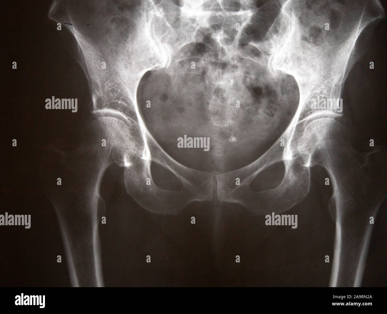
Xray Of A Human Pelvis Stock Photo Alamy from c8.alamy.com Ct, us, and mr provide comprehensive evaluation of the abdomen including the peritoneal cavity, retroperitoneal compartments, abdominal and pelvic organs, blood vessels, and lymph nodes. Use the mouse scroll wheel to move the images up and down alternatively use the tiny arrows (>>) on both side of the image to move the images.>>) on both side of the image to move the images. Imaging evaluation to determine the status of pelvic lymph nodes has yet to attain benchmark status clinically. Hover on/off image to show/hide findings. Systematic review three rings trace the main pelvic ring and two obturator foramina if a ring is disrupted, think fracture. For more anatomy content please follow us and visit our website: The routine pelvic view is anteroposterior (ap) projection, and in 94% of cases, a correct diagnosis can be made from this view. A pelvic ultrasound is a noninvasive diagnostic exam that produces images that are used to assess organs and structures within the female pelvis.
This article provides an overview of normal anatomy of the pelvic floor as seen on magnetic resonance imaging, ultrasound, and fluoroscopic studies performed in.
On the other hand, current imaging techniques still have the potential to improve the detection of micrometastatic lesions and differentiation between hyperplastic and. In an adult, the innominate bones consist of the fused ilium, ischium, and pubis (figure 1). Anatomy of the pelvis the pelvis is a ring of bones situated between the spine and the legs. On one hand, pelvic lymphatic drainage is complex and varies depending on the types and locations of primary tumors. Annotated case courtesy of dr phillip marsh, radiopaedia.org, rid: For more anatomy content please follow us and visit our website: Pelvis anatomy the pelvis is either the lower part of the trunk of the human body between the abdomen and the thighs. Until puberty, each hip bone consists of three separate bones yet to be fused: Ct of the pelvis is the technique of choice for evaluating complex fracture patterns, degree of displacement and soft tissue injury. Quizzes about radiology anatomy quiz. Knowledge of normal pelvic floor anatomy can aid the radiologist in understanding the complex nature of pelvic floor dysfunction and is important for comprehensive image interpretation. The routine pelvic view is anteroposterior (ap) projection, and in 94% of cases, a correct diagnosis can be made from this view. Tap on/off image to show/hide findings.
Annotated case courtesy of dr phillip marsh, radiopaediaorg, rid: pelvic anatomy. This mri male pelvis axial cross sectional anatomy tool is absolutely free to use.