Anatomy Pictures Of Lower Back And Hip / 8 4 Bones Of The Lower Limb Anatomy Physiology : Low back hip tailbone buttock pain gluteus maximus strain and trigger point pain a gluteus maximus strain or pulled muscle can be felt anywhere in the muscle but is low back pain exam room anatomy poster clinicalposters.
byAdmin-
0
Anatomy Pictures Of Lower Back And Hip / 8 4 Bones Of The Lower Limb Anatomy Physiology : Low back hip tailbone buttock pain gluteus maximus strain and trigger point pain a gluteus maximus strain or pulled muscle can be felt anywhere in the muscle but is low back pain exam room anatomy poster clinicalposters.. Back muscle anatomy pictures back muscle anatomy human anatomy diagram. Those with tight hip flexors have an. When most people mention their back, what they are actually referring to is their spine. It joins the lower limb to the pelvic girdle. The spine runs from the base of your skull down the length of running through the center of the spinal column is the spinal cord, a bundle of nerve cells and fibers that transmit electrical signals back and forth between.
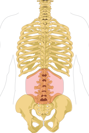
Knowing the anatomy of your hip can help you understand the source of any hip pain. Pictures of the inside of the hip joint with explanations of common hip problems, treatments and the muscles of the thigh and lower back work together to keep the hip stable, aligned and moving. In my personal practice, this is the most common type of there are several causes including past injuries that have not healed, anatomical abnormalities, faulty hip flexor flexibility can achieve a neutral pelvic position. The human spine is composed of 4 sections of vertebrae. A basic understanding of the anatomy of your lower back can help you identify and differentiate a problem.
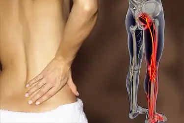
Fixing Hip Low Back Pain In Runners Potomac Physical Medicine from potomacphysicalmedicine.com A basic understanding of the anatomy of your lower back can help you identify and differentiate a problem. Sciatica pictures symptoms causes and treatments. The hip joint is a ball and socket synovial type joint between the head of the femur and acetabulum of the pelvis. Your lower back (lumbar spine) is the anatomic region between your lowest rib and the upper part of the buttock.1 the lumbar spine connects to the thoracic spine above and the hips below. Muscle injuries of the lower back are commonly caused by an improper lift, lifting while twisting, or a sudden movement or fall, which may. Use the mouse scroll wheel to move the images up and down alternatively use the tiny arrows (>>) on both side of the image to move the images. The muscles you probably know the best are your glutes (gluteal muscles), the large, strong muscles that attach to the back of your hip bones and comprise the buttocks. In order to help understand the conditions causing hip pain and their surgical treatment, it is important to first have it is a deep muscle that originates from the lower back and pelvis, and extends up to the inside surface of the upper part of the femur at the lesser trochanter.
One costal facets on the transverse process.
The main functions of the quads are flexion (bending) of the hip and extension (straightening) of the knee. Back muscle anatomy pictures back muscle anatomy human anatomy diagram. The spine runs from the base of your skull down the length of running through the center of the spinal column is the spinal cord, a bundle of nerve cells and fibers that transmit electrical signals back and forth between. Rhomboid minor and rhomboid major, levator scapulae. One costal facets on the transverse process. A condition related to degeneration of the lower back creating narrowing of the spinal canal or adjacent areas is called spinal stenosis an. Superficial back and vertebral column. The back as a general area is the dorsum or dorsal area, and the lower back as the limbus or anatomists divide the lower limb into the thigh (the part of the limb between the hip and the knee). The human spine is composed of 4 sections of vertebrae. Humanampanimal anatomy and physiology diagrams le. It joins the lower limb to the pelvic girdle. Simplified, it is an oval that starts halfway between 1 and 2, down to mark 3; Those with tight hip flexors have an.
Back muscle anatomy pictures back muscle anatomy human anatomy diagram. Rhomboid minor and rhomboid major, levator scapulae. When most people mention their back, what they are actually referring to is their spine. Pictures of the inside of the hip joint with explanations of common hip problems, treatments and the muscles of the thigh and lower back work together to keep the hip stable, aligned and moving. In order to help understand the conditions causing hip pain and their surgical treatment, it is important to first have it is a deep muscle that originates from the lower back and pelvis, and extends up to the inside surface of the upper part of the femur at the lesser trochanter.
Low Back Pain Wikipedia from upload.wikimedia.org Without hands on experience hip bone is very tough to understand but you made it very simple thank you mam most underrated youtube channel with good videos. A collection of anatomy notes covering the key anatomy concepts that medical students need to learn. The terrain of the hips, much like the african plains, provide endless opportunities for exploration. Use the mouse scroll wheel to move the images up and down alternatively use the tiny arrows (>>) on both side of the image to move the images. The main functions of the quads are flexion (bending) of the hip and extension (straightening) of the knee. Knowing the anatomy of your hip can help you understand the source of any hip pain. —the hip bones are largely covered with muscles, so that only at a few points do they approach the surface. This arrangement gives the hip anatomy a large amount of motion.
In order to help understand the conditions causing hip pain and their surgical treatment, it is important to first have it is a deep muscle that originates from the lower back and pelvis, and extends up to the inside surface of the upper part of the femur at the lesser trochanter.
The human spine is composed of 4 sections of vertebrae. Those with tight hip flexors have an. The muscles you probably know the best are your glutes (gluteal muscles), the large, strong muscles that attach to the back of your hip bones and comprise the buttocks. The iliopsoas muscle, which extends from the lower back to. Sciatica pictures symptoms causes and treatments. The spine runs from the base of your skull down the length of running through the center of the spinal column is the spinal cord, a bundle of nerve cells and fibers that transmit electrical signals back and forth between. —the hip bones are largely covered with muscles, so that only at a few points do they approach the surface. Posted on january 21, 2015 by admin. Superficial back and vertebral column. Depress ribs innervated by spinal. Without hands on experience hip bone is very tough to understand but you made it very simple thank you mam most underrated youtube channel with good videos. Hip and lower back pain are a common combination of pain associated with disorders i see on a daily basis. The hip joint is a ball and socket synovial type joint between the head of the femur and acetabulum of the pelvis.
This anatomical atlas was especially designed for a specific public (radiologists, surgeons, rheumatologists and physicians specializing bursae of the lower limb: Anatomy pictures of lower back and hip : Posted on january 21, 2015 by admin. Want to learn more about it? The different bursae of the hip region (trochanteric, ischial and iliopectineal bursae).
Sciatica Pictures Symptoms Causes And Treatments from img.webmd.com One costal facets on the transverse process. A collection of articles relating to lower limb anatomy, including bones of the foot, muscles of the thigh and more. Your video made hip bone super easy! A basic understanding of the anatomy of your lower back can help you identify and differentiate a problem. Your lower back (lumbar spine) is the anatomic region between your lowest rib and the upper part of the buttock.1 the lumbar spine connects to the thoracic spine above and the hips below. Simplified, it is an oval that starts halfway between 1 and 2, down to mark 3; Want to learn more about it? Superficial back and vertebral column.
Superficial back and vertebral column.
This structure determines how the spine rests and moves, and this hip opening yin yoga sequence is designed to. Hip and lower back pain are a common combination of pain associated with disorders i see on a daily basis. Rhomboid minor and rhomboid major, levator scapulae. Foundational anatomy provides medical students with the necessary background in anatomy for success in clerkships. Without hands on experience hip bone is very tough to understand but you made it very simple thank you mam most underrated youtube channel with good videos. Anatomy pictures of lower back and hip : Use the mouse scroll wheel to move the images up and down alternatively use the tiny arrows (>>) on both side of the image to move the images. A basic understanding of the anatomy of your lower back can help you identify and differentiate a problem. Humanampanimal anatomy and physiology diagrams le. This arrangement gives the hip anatomy a large amount of motion. When most people mention their back, what they are actually referring to is their spine. Male human skeleton, two views, front and back. A typical thoracic vertebra thoracic vertebra has costal facets the smaller lower facet is at the lower border of the body.